
OME: A Comprehensive Guide to Otitis Media with Effusion for Healthcare Students (2026)
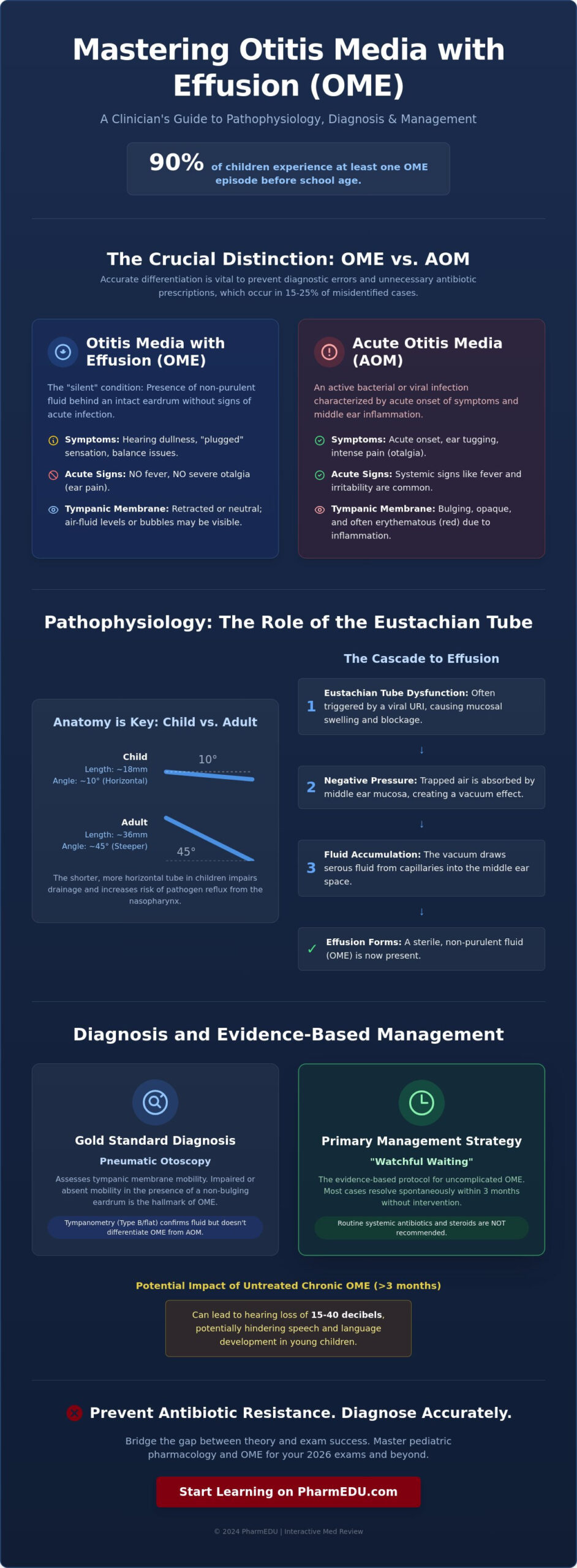
Could the most effective pharmacological intervention for a middle ear effusion actually be no medication at all? While your instinct might be to treat every pediatric ear complaint with urgency, clinical research indicates that nearly 90% of children experience at least one episode of ome before they reach school age. You’ve likely felt the stress of clinical rotations where the distinction between a red, bulging drum and a simple fluid buildup feels ambiguous. It’s common to struggle with the exact timing for antibiotic intervention or to feel overwhelmed by the latest pediatric treatment protocols during a busy shift.
This guide provides the precision you need to master the pathophysiology, diagnosis, and pharmacological management of OME for your 2026 exams and future practice. You’ll gain a board-ready understanding of how to differentiate OME from acute otitis media with total confidence. We’ll examine the specific criteria for the “watchful waiting” strategy and provide a structured framework for managing pediatric ear health that aligns with the highest professional standards.
Key Takeaways
- Learn to accurately distinguish between middle ear fluid and acute infection to prevent common diagnostic errors in clinical practice.
- Understand how Eustachian tube dysfunction and negative middle ear pressure drive the underlying pathophysiology of ome.
- Identify hallmark symptoms like hearing loss and master pneumatic otoscopy as the gold standard for a definitive diagnosis.
- Master the “Watchful Waiting” protocol and the evidence-based rationale for avoiding routine systemic antibiotics in management.
- Leverage high-yield video vignettes and interactive flashcards to bridge the gap between pharmacological theory and exam success.
Understanding OME: Definition and the Crucial Difference from AOM
Otitis media with effusion, commonly referred to as ome, represents one of the most frequent reasons for pediatric office visits and elective surgery in developed nations. Unlike other forms of Otitis media, this condition doesn’t typically present with the dramatic symptoms clinicians associate with ear infections. It’s often dubbed a “silent” condition because patients, particularly young children, rarely complain of the sharp pain or high fevers that characterize acute episodes. Data from the American Academy of Pediatrics indicates that 90% of children experience at least one episode of effusion before age five, often following an upper respiratory infection. This high prevalence makes it a cornerstone of pediatric otolaryngology, especially considering its impact on hearing development. A 2022 study published in the Journal of Otolaryngology found that persistent effusion lasting longer than three months is associated with measurable delays in phonological processing.
The Clinical Definition of OME
Clinical identification centers on the presence of non-purulent, often serous or mucoid fluid within the middle ear space behind an intact tympanic membrane. Clinicians won’t find the systemic markers of infection here; fever, severe otalgia, and irritability are notably absent. The fluid acts as a physical barrier to sound conduction, potentially leading to a hearing loss of 15 to 40 decibels. This deficit can significantly hinder speech and language development during critical windows of neuroplasticity. OME is a chronic inflammatory state rather than an acute infection.
OME vs. AOM: A Comparative Framework
Accurate differentiation between ome and Acute Otitis Media (AOM) is a vital skill for preventing diagnostic errors and antibiotic resistance. AOM presents with a bulging, opaque, and often erythematous tympanic membrane, indicating an active bacterial or viral process. In contrast, an ear with effusion might show a retracted or neutral membrane with visible air-fluid levels or bubbles. Misidentifying OME as AOM leads to unnecessary antibiotic prescriptions in approximately 15% to 25% of pediatric cases. Key differentiators include:
- AOM: Involves acute onset of symptoms like ear tugging, intense pain, and fever.
- OME: Presents with hearing dullness, a “plugged” sensation, or no symptoms at all.
- Diagnostic Tools: Tympanometry serves as a critical diagnostic tool, where a Type B (flat) tympanogram confirms the presence of fluid but doesn’t distinguish infection from sterile fluid.
Clinicians must rely on pneumatic otoscopy to assess membrane mobility, which remains the gold standard for differentiating these two states. Proper identification ensures that patients receive appropriate monitoring instead of ineffective antimicrobial therapy. This distinction is the first step in providing evidence-based care that aligns with modern clinical guidelines for 2026.
The Pathophysiology of Otitis Media with Effusion
The development of ome centers on the failure of the Eustachian tube to maintain a sterile, air-filled middle ear environment. Under healthy conditions, this tube opens intermittently during swallowing or yawning to equalize pressure and drain secretions. When this mechanism fails, the middle ear transforms into a closed system. This dysfunction isn’t just a mechanical blockage; it’s a complex failure of pressure regulation and mucosal health that sets the stage for fluid accumulation.
Eustachian Tube Mechanics and Fluid Dynamics
Anatomical differences play a decisive role in why certain populations are more vulnerable. In pediatric patients, the Eustachian tube is roughly 18mm long and sits at a 10-degree angle, making it nearly horizontal. By adulthood, this structure matures to approximately 36mm in length at a steeper 45-degree angle. This horizontal orientation in children significantly increases the risk of retrograde reflux of nasopharyngeal pathogens into the middle ear. According to the Otitis Media with Effusion (OME) research from the Children’s Hospital of Philadelphia, viral upper respiratory infections often trigger the initial mucosal edema. This swelling obstructs the tube, causing the middle ear mucosa to absorb trapped air. The resulting negative pressure creates a vacuum effect, drawing serous fluid from the surrounding capillaries into the tympanic cavity. Environmental triggers, such as secondhand smoke, worsen this state by paralyzing the cilia responsible for clearing these fluids.
Inflammatory Mediators and Biofilms
Chronic cases of ome often persist long after the initial viral trigger has resolved. This persistence is frequently driven by a cocktail of inflammatory mediators, including Interleukin-1 and Tumor Necrosis Factor-alpha, which increase the permeability of local blood vessels and stimulate mucus-secreting glands. Clinical data from 2023 indicates that bacterial biofilms are present in up to 92% of chronic middle ear effusions. These complex microbial communities act as a protective shield, making bacteria highly resistant to both the host’s immune response and standard antibiotic treatments. Because traditional antibiotics typically target actively dividing planktonic bacteria, the dormant cells tucked within a biofilm remain untouched. This explains why standard pharmacological interventions often fail in chronic cases, necessitating surgical options like tympanostomy tubes. Understanding these molecular shifts is vital for modern clinical practice. Healthcare students can deepen their expertise by exploring accredited courses on pediatric pathology to stay current with these evolving diagnostic challenges.
Clinical Presentation and Diagnostic Criteria for OME
Unlike acute otitis media (AOM), OME typically lacks systemic signs of infection such as fever or severe otalgia. Patients often present with a “plugged” sensation or mild discomfort rather than sharp pain. In pediatric populations, caregivers might report that the child has become less responsive to verbal cues or has increased the volume on electronic devices. Clinical data suggests that Otitis media with effusion can result in a conductive hearing loss of 25 to 30 decibels, which significantly impacts auditory processing during early development.
Healthcare providers frequently identify OME during routine wellness checks or when evaluating unrelated upper respiratory complaints. Because the condition is often painless, it can persist unnoticed for weeks. If the effusion remains for more than 90 days, clinicians classify it as chronic OME. This prolonged duration increases the risk for speech and language delays, particularly in children under age 3 who are in critical developmental windows. Balance issues or general clumsiness are also reported, as the fluid affects middle ear pressure and vestibular function. Identifying these subtle signs early is vital for preventing long-term educational hurdles.
Physical Examination Findings
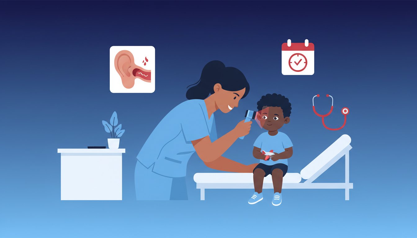
Pneumatic otoscopy remains the gold standard for clinical diagnosis in any setting. During the exam, the healthcare provider observes the tympanic membrane (TM) for specific markers. In OME, the TM often appears in a neutral or retracted position rather than bulging. You might see a yellowish, amber, or even bluish fluid behind the drum. Air-fluid levels or visible bubbles are pathognomonic signs. The defining characteristic is limited or absent mobility of the drum when applying pressure with the pneumatic bulb. If the drum doesn’t move, it’s a strong indicator of middle ear fluid.
Diagnostic Testing: Tympanometry and Audiometry
Objective testing provides essential data to support the physical exam findings. Tympanometry measures the compliance of the middle ear system and the pressure within the ear canal. A “Type B” tympanogram, characterized by a flat tracing with no identifiable peak, indicates the presence of fluid. When ome persists for 3 months or longer, or if there’s a suspected developmental delay, you must refer the patient for a full audiologic evaluation. This assessment determines the exact degree of hearing loss and guides the decision-making process for surgical interventions like myringotomy tubes. Regular screening ensures that “silent” cases don’t go untreated for extended periods.
Pharmacological Management and Watchful Waiting Strategies
Effective clinical management of ome centers on the “Watchful Waiting” protocol. The American Academy of Pediatrics (AAP) clinical practice guidelines recommend a three-month observation period from the date of effusion onset or initial diagnosis. This conservative approach recognizes that the majority of cases are self-limiting. Healthcare providers must monitor hearing levels and middle ear status during this window instead of immediately prescribing medication. It’s a period of active surveillance where the clinician educates the family on the natural history of the condition and the low risk of permanent damage during this timeframe.
The Argument Against Routine Antibiotics
Systemic antibiotics aren’t recommended for routine management because they don’t address the underlying Eustachian tube dysfunction. Clinical trials show that 75-90% of ome cases resolve within three months without intervention. Using antibiotics creates unnecessary risks, such as gastrointestinal distress and the development of antimicrobial resistance, for a marginal short-term benefit. Data suggests that the rate of spontaneous resolution following an upper respiratory infection is high enough that the potential side effects of drugs like amoxicillin outweigh their clinical utility. Evidence-based practice dictates that we reserve these agents for confirmed bacterial infections rather than sterile effusions.
The Role of Intranasal Corticosteroids and Decongestants
Decongestants don’t facilitate fluid drainage in the middle ear. These medications target the nasal mucosa but fail to reach the Eustachian tube effectively. Similarly, antihistamines show no benefit unless the patient has documented allergic rhinitis. While some studies investigated intranasal corticosteroids for reducing adenoidal inflammation, the evidence remains conflicting and largely points to a lack of efficacy in clearing middle ear fluid. Over-medicating pediatric patients can lead to avoidable side effects, including epistaxis, sleep disturbances, and increased heart rate. Practitioners should prioritize evidence-based observation over polypharmacy.
When to Consider Surgical Intervention (Tympanostomy Tubes)
Surgical referral is the standard of care when the effusion persists for three months or longer and causes significant morbidity. The primary criteria for surgery include a documented hearing loss threshold of 20 decibels or worse in the better ear. Tympanostomy tubes serve as a pressure equalization mechanism, allowing the middle ear to stay ventilated while the Eustachian tube recovers. Post-surgical care often involves the use of otic antibiotic drops, such as ofloxacin, to prevent postoperative otorrhea or tube blockage. This targeted pharmacological approach is far more effective than systemic treatments for maintaining tube patency.
Master Pediatric Pharmacology and OME with PharmEDU
Transitioning from classroom theory to clinical application requires more than just reading textbooks. PharmEDU bridges this gap with high-yield video vignettes that specifically target pediatric ear pathologies. These visual tools allow you to see the physical manifestations of ome alongside expert commentary, which is vital for developing diagnostic intuition. To ensure you don’t forget the nuances of treatment, our interactive flashcards use spaced repetition to help you memorize complex antibiotic dosing and surgical referral guidelines.
Distinguishing between Otitis Media with Effusion and Acute Otitis Media (AOM) is a frequent challenge during 2026 clinical rotations. Our platform provides detailed case studies that force you to analyze tympanometry and otoscopy findings in real-time. By engaging with these scenarios, you’ll learn to recognize the “glue ear” appearance of ome without the signs of acute infection. PharmEDU stands as the definitive resource for pharmacology mastery, providing the precision you need to excel in your medical or pharmacy career.
- High-yield videos covering pediatric ENT pathologies and tympanic membrane assessment.
- Interactive flashcards designed for long-term retention of drug mechanisms and side effects.
- Clinical simulations that differentiate chronic effusion from acute bacterial infections.
Board Review for OME and Pediatric Pathologies
Preparing for the NAPLEX or NCLEX requires a deep understanding of pediatric topics, where even small dosing errors have serious consequences. We’ve simplified the process by creating a pharmacology study guide for nursing students that breaks down drug classes into manageable segments. Research shows that micro-learning modules lasting 8 to 12 minutes increase retention by up to 20 percent compared to traditional long-form lectures. Our platform delivers these bite-sized lessons, making it easy to study during a short break or between clinical shifts.
Join the PharmEDU Community for Professional Success
Gain full access to our extensive pharmacology library by becoming a member of the PharmEDU community. We’ve optimized every resource for mobile devices, so you can review pediatric guidelines on the go. Whether you’re in the university library or on the hospital ward, professional growth is always within reach. Our digital mentor approach ensures you’re never alone when facing complex medical regulations or new clinical findings. Start your PharmEDU subscription today to secure your academic success and prepare for your professional future with confidence.
Advancing Your Clinical Expertise in Pediatric Otology
Mastering the distinction between acute infection and fluid accumulation helps prevent the 30% of unnecessary antibiotic prescriptions often recorded in pediatric primary care. Effective management of ome relies on adhering to the 3-month watchful waiting period established by clinical practice guidelines to avoid premature surgical intervention. Students who prioritize these diagnostic criteria and pharmacological nuances provide safer, more effective care in their future clinical practices. It’s about bridging the gap between textbook theory and real-world patient outcomes.
Navigating the complexities of medical school and board preparation doesn’t have to be an overwhelming administrative burden. PharmEDU acts as your digital mentor, offering expert-curated content that simplifies even the most difficult topics. You’ll benefit from high-yield video vignettes for rapid learning and interactive flashcards that help you master active recall techniques. With over 1,000 peer-reviewed quizzes, you can track your progress and ensure you’re ready for every clinical challenge. Our platform ensures you’re always aligned with the latest evidence-based standards.
Master Pharmacology with PharmEDU Subscriptions
Your journey toward becoming a top-tier healthcare provider is just beginning, and we’re here to support your professional growth every step of the way.
Frequently Asked Questions
What is the primary difference between OME and AOM?
The primary difference lies in the absence of acute inflammatory symptoms like fever or severe otalgia in OME compared to Acute Otitis Media (AOM). AOM is characterized by a rapid onset of pain and signs of infection, whereas ome involves fluid accumulation in the middle ear without these systemic signs. Clinical studies show that 40% of children still have residual fluid four weeks after an AOM episode, which then transitions into this non-infectious state.
Do antibiotics help clear the fluid in OME?
Antibiotics aren’t effective for clearing middle ear fluid because ome isn’t an active bacterial infection. Clinical trials demonstrate that the rate of resolution doesn’t significantly improve with antimicrobial therapy compared to a watchful waiting approach. The American Academy of Otolaryngology guidelines specifically recommend against using antibiotics for this condition unless a concurrent infection is present. This approach avoids unnecessary side effects and helps prevent antibiotic resistance in pediatric populations.
How long does Otitis Media with Effusion typically last?
Most cases of Otitis Media with Effusion resolve spontaneously within 12 weeks without medical intervention. Research indicates that 75% to 90% of children see their symptoms clear within this three-month window. If the fluid persists beyond 90 days, the condition’s classified as chronic. At this stage, the likelihood of spontaneous resolution drops to approximately 20% to 30%, which requires more frequent clinical monitoring by healthcare providers.
Can OME cause permanent hearing loss in children?
Permanent hearing loss is rare, but the condition frequently causes a temporary conductive hearing loss averaging 25 to 30 decibels. This occurs because the fluid prevents the ossicles and tympanic membrane from vibrating correctly. Long-term structural damage like cholesteatoma or tympanosclerosis can occur in less than 5% of chronic cases if the ear’s left unmonitored. Early detection ensures these complications don’t lead to irreversible auditory deficits or developmental delays.
Why are decongestants not recommended for OME treatment?
Decongestants aren’t recommended because they don’t address the underlying Eustachian tube dysfunction responsible for the fluid. Multiple clinical trials and Cochrane reviews show that these medications fail to improve resolution rates or reduce the need for surgery. Since they don’t reach the middle ear effectively, their use only exposes the patient to potential side effects like irritability or increased heart rate. They provide no measurable clinical benefit for middle ear clearance.
When should a child with OME be referred to an ENT specialist?
A child should be referred to an ENT specialist if the fluid persists for 3 months or if there’s a documented hearing loss of 20 decibels or more. Referral’s also necessary if the child shows signs of speech delay or structural changes to the eardrum. Specialists typically consider surgical options, like tympanostomy tubes, for children who meet these specific criteria. This intervention helps prevent developmental setbacks and improves the child’s quality of life.
Is OME contagious between children?
Otitis Media with Effusion isn’t contagious and can’t be spread from person to person. While the initial upper respiratory infection that caused the Eustachian tube blockage might be viral and transmissible, the resulting middle ear fluid is sterile or contains non-replicating bacteria. Children with this condition don’t need to be isolated from school or daycare settings as long as they don’t have an active fever. It’s a localized mechanical issue rather than a communicable disease.
What does a flat tympanogram (Type B) indicate in a clinical setting?
A Type B tympanogram indicates a lack of tympanic membrane mobility, which strongly suggests the presence of middle ear fluid. This flat tracing shows that the eardrum can’t move in response to pressure changes because of the liquid trapped behind it. In a clinical setting, a Type B result with a normal ear canal volume, typically 0.3 to 1.0 ml in children, has a 90% predictive value for effusion. It’s a vital diagnostic tool for confirming clinical findings.